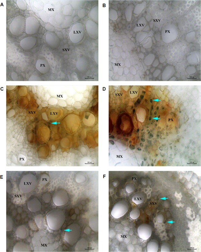
Figure 3.
Bright field microscopy root images showing high fungal colonization in xylem regions of plants under combined stress. Transverse hand sections of plant roots from individual and combined stress treatments were stained with lactophenol aniline blue and observed under 40X objective with fixed 40X condenser of LMI BM-X microscope. No fungal mycelia were observed in the xylem of control (A) and drought (B) treated plants. Plant roots treated with F. solani infection have less number of stained mycelia (C) whereas plants treated with combined drought and F. solani infection showed large number of stained mycelia (D). Plant roots with R. bataticola infection (pathogen only) showed less stained mycelia (E) whereas plants treated with combined drought and R. bataticola showed large number of stained mycelia (F). The cyan arrow indicates the aniline blue stained fungal mycelia. Scale bar represents 150 µm, microscopy was done with three technical replicates. Two independent experiments showed similar result. MX, metaxylem; PX, protoxylem; LXV, large xylem vessel; SXV, small xylem vessel; dotted line demarcates the metaxylem and protoxylem. Note: the blue arrows are for pointing out stained fungal mycelia and not for quantification. Further, number of stained mycelial structures were counted under each microscopic field and the values are 20 ± 6 (C), 94 ± 5 (D),19 ± 2 (E) and 53 ± 5 (F). Besides blue staining, the dark brown colour is due to infection (see Supplementary Fig. 23). The difference in diameter of xylem vessels between control and drought could be due to treatment effect. All the images are captured under white balance background.

