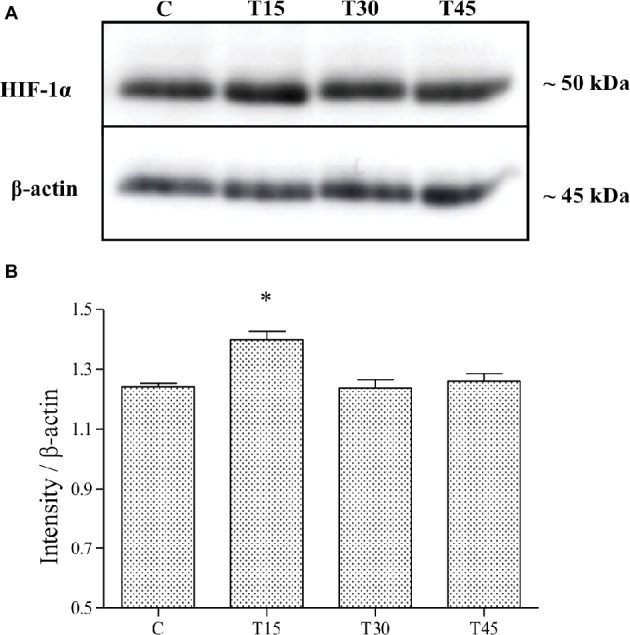
Figure 3.

HIF-1α expression was evaluated by Western blotting analysis (A) in hearts from trained and control mice and its intensity measured with an image software (B). HIF-1α levels resulted significantly higher in T15 than in C, T30, and T45 groups (*p < 0.05, T15 vs. C, T30, and T45).
