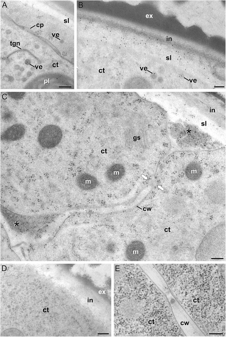
FIGURE 3.
JIM13 immunogold labeling. (A–C): Images of dividing embryogenic microspores, showing details of a maturing cell plate (cp) in (A), the intine (in) and subintinal layer (sl) in (B), and a fragmented cell wall (cw) with gaps (white arrows) and deposits of cytoplasmic material (asterisks) secreted to the apoplast in (C). (D) Pollen-like structure. (E) Torpedo MDE. ct: cytoplasm; ex: exine; gs: Golgi stack; m: mitochondrion; pl: plastid; tgn: trans-Golgi network; ve: vesicle. Bars: 100 nm.

