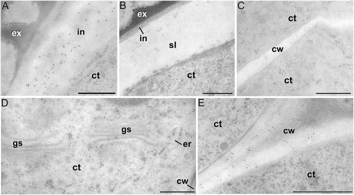
FIGURE 8.
Xyloglucan immunogold labeling with the mAb CCRC-M1. (A) Detail of the intine (in) in a pollen-like structure. (B) Detail of the intine and subintinal layer (sl) of an embryogenic microspore. Note that the intine wall is strongly labeled in pollen-like structures (A), but not in embryogenic microspores (B). (C) Inner cell wall (cw) of an embryogenic microspore. (D,E) Torpedo MDEs. Details of the cytoplasm (ct) are shown in (D), where cytoplasmic gold particles strongly decorate the Golgi stacks (gs) and associated vesicles. The cell wall is shown in (E). ex: exine; er: endoplasmic reticulum. Bars: 500 nm.

