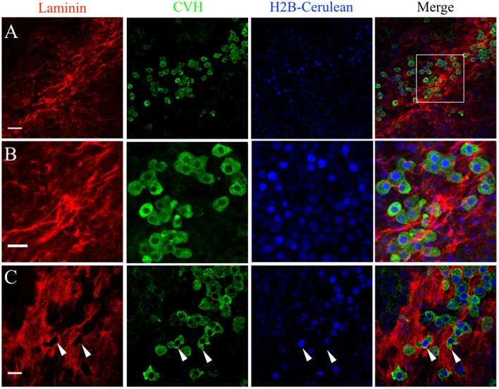
Figure 6.
PGCs in the germinal crescent are intimately associated with the laminin fibril meshwork. Whole-mount immunofluorescence for laminin (ab31-2) and CVH from two locations in the lateral germinal crescent of an HH5 [Tg(hUbC:H2B-Cerulean-2A-Dendra2)] quail embryo. Images in (A) are single optical sections (3.2 μm Z depth) taken at a lateral region of the germinal crescent using a 20x 0.8NA objective. Scale bar = 50 um. The bounding box in the merge indicates the region shown in (B) at increased magnification. Scale bar = 25 μm. (C) Images are maximum intensity projections of 3 optical sections (2.3 μm each) from 40x confocal Z stacks taken from the dorsal aspect of the embryo at the lateral germinal crescent. White arrows in (C) highlight regions of laminin that appear completely displaced by a PGC. Scale bar = 25 μm.

