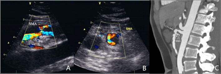
Figure 1.
Type I SISMAD. Representative images of a 48 year-old male are provided. The patient was admitted due to sudden abdominal pain for 4 h and was diagnosed with SISMAD by CTA and color Doppler sonography. (A) Blood flow and perforation between true and false lumen on long axis CDS imaging of the SMA revealed the entry site and thrombosis in the false lumen. (B) Blood flow and perforation between true and false lumen on short axis cross-section CDS imaging of the SMA revealing less blood flow and thrombosis in the false lumen. (C) CTA image of type I lesion. SISMAD, spontaneous isolated superior mesenteric artery dissection; CTA, computed tomography angiography.

