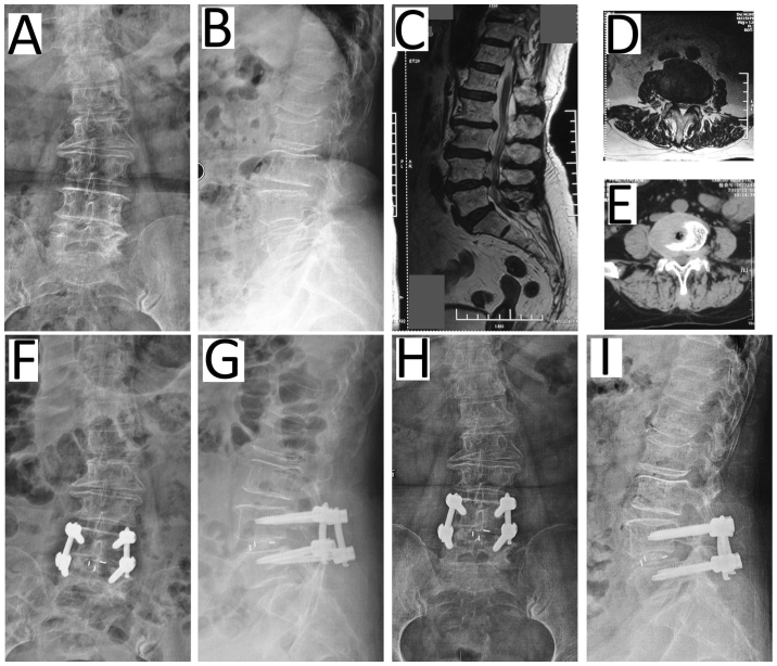
Figure 2.
Representative patient (female, 58 years) with degenerative disk disease of L4/5 undergoing c-TLIF. Preoperative (A) anteroposterior and (B) lateral radiographs and (C) the MRI scan highlighting the herniated disc of L4/5. The herniated disc was on the lateral right on (D) MRI and (E) computed tomography scans. At 7 days post c-TLIF with pedicle screw fixation, (F) anteroposterior and (G) lateral radiographs exhibited the restoration of the disc space height. (H) Anteroposterior and (I) lateral radiographs recorded 12 months post surgery. c-TLIF, classical open transforaminal lumbar interbody fusion; MRI, magnetic resonance imaging.

