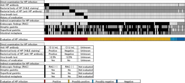
Figure 1.

Evaluation for Helicobacter pylori (HP) infection. Infection was evaluated directly and indirectly (top panel), and color coded using the criteria indicated in the middle panel. Patients positive for any direct examination were grouped as “positive” (red), and positive for typical endoscopic findings, (ie, absence of regular arrangement of collecting venules [RAC]) were “possibly positive” (yellow). The remaining patients were considered as “possibly negative” (light blue) or “negative” (dark blue)
