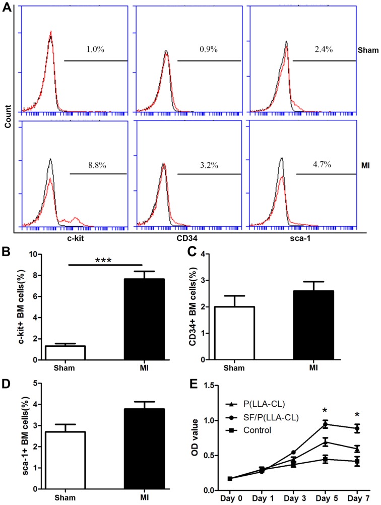
Figure 3.
Flow cytometric analysis and proliferation assay. (A) Flow cytometric analysis plots of c-kit+, CD34+ and sca-1+ cells isolated from MI and sham mice. (B-D) Expression analysis of c-kit+, CD34+ and sca-1+ in BM cells. ***P<0.001 as indicated (n=6 for each group). (E) CellTiter 96® AQueous One Solution reagent was used to assess c-kit+ BM cells seeded on P(LLA-CL) or SF/P(LLA-CL) or those cultured without scaffolds at indicated time points. *P<0.05 (n=6 for each group). SF, silk fibroin; P(LLA-CL), poly(L-lactic acid-co-ε-caprolactone); MI, myocardial infarction; BM, bone marrow; CD, cluster of differentiation; CD117, c-kit; sca-1, stem cell antigen-1; OD, optical density.

