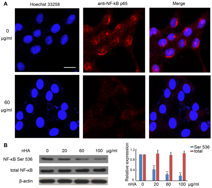
Figure 5.
nHA inhibited NF-κB p65 nuclear translocation and protein expression. (A) Images of fluorescent staining with Hoechst 33258 and anti-NF-κB p65 antibody. C6 cells were treated with 60 µg/ml nHA for 24 h and stained with Hoechst 33258 and anti-NF-κB p65 antibody. Images (original magnification, ×20) were captured using a confocal fluorescent microscope. Experiments were repeated at least 3 times and representative images are presented. The scale bar represents 15 µm. (B) Images of western blotting analysis for NF-κB p65 (Ser 536) and total NF-κB p65 in C6 cells that were treated with 0, 20, 60 or 100 µg/ml for 24 h. β-actin was used as a loading control. The experiment was repeated 3 times. The relative expression of NF-κB was analyzed. **P<0.01 vs. 0 µg/ml nHA. nHA, nano-hydroxyapatite; NF, nuclear factor.

