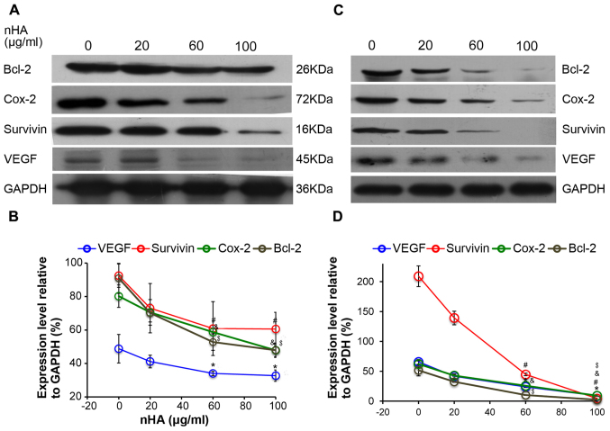
Figure 6.
nHA reduced protein expression of nuclear factor-κB p65 target molecules. (A) Representative images of western blotting of C6 cells. (B) Densitometry analysis of the western blotting of C6 cells. (C) Representative images of western blotting of U87MG ATCC cells. (D) Densitometry analysis of the western blotting of U87MG ATCC cells. C6 and U87MG ATCC cells were treated with 0, 20, 60 or 100 µg/ml nHA for 24 h. The experiment was repeated 3 times. Representative images are presented. *P<0.05, VEGF expression vs. 0 group; #P<0.05, Survivin expression vs. 0 group; &P<0.05, Cox-2 expression vs. 0 group; $P<0.05, Bcl-2 expression vs. with 0 group. nHA, nano-hydroxyapatite; Bcl-2, B cell lymphoma-2; Cox-2, cyclooxygenase-2; VEGF, vascular endothelial growth factor.

