Figure 1.

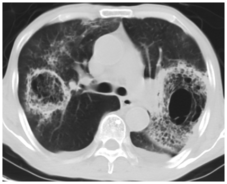
CT image of lung with pulmonary mucormycosis. The bilateral lungs show a symmetrical distribution of glass-like shadows with blurry boundaries and patchy solid shadows. CT, computed tomography.

CT image of lung with pulmonary mucormycosis. The bilateral lungs show a symmetrical distribution of glass-like shadows with blurry boundaries and patchy solid shadows. CT, computed tomography.