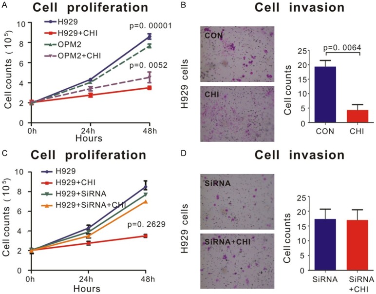
Figure 3.

Chidamide inhibited proliferation and invasion of MM cells via SDHA. A. OPM2 and H929 cells lines treated by 6 μM chidamide or DMSO for 48 h. Cell proliferation was measured as described in Materials and Methods. Cells treated by chidamide proliferated much slower than cells treated by DMSO, especially in H929 cells. B. By transwell invasion assay, invasion ability of H929 cells was significantly inhibited by 6 μM chidamide treatment than that by DMSO. Quantifications of cell invasion were shown in the right panel. C. SDHA siRNA and control siRNA were used as described in Materials and Methods. The ability of proliferation of H929 cells transfected with SDHA siRNA remained a same level no matter treated by chidamide or DMSO. D. By transwell invasion assay, SDHA siRNA transfected cells acquired no decrease in cell invasion after chidamide treatment. Quantifications of cell invasion were shown in the right panel.
