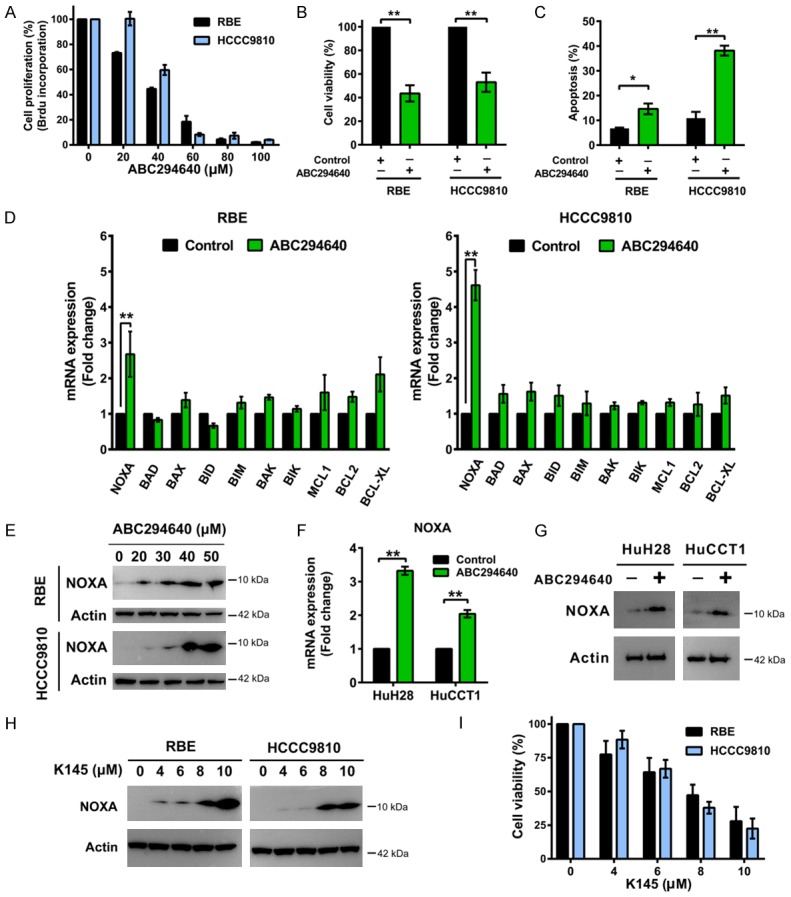
Figure 1.

SPHK2 inhibition suppresses cholangiocarcinoma cell growth, induces apoptosis and upregulates NOXA expression. A. RBE and HCCC9810 cells were treated with ABC294640 for 72 h and cell proliferation was quantified by BrdU ELISA assay. B. Cells were treated with ABC294640 at 50 μM for 72 h and cell viability was determined by CCK-8 assay. C. Cells were treated with ABC294640 at 50 μM for 72 h and cell apoptosis was then measured by Annexin V-FITC/PI labeling followed by flow cytometry. D. Real-time qPCR analysis of BCL2 family mRNA level in RBE and HCCC9810 cells treated with 50 μM ABC294640 or no drug control for 24 h. E. Western immunoblotting analysis of NOXA protein levels in RBE and HCCC9810 cells treated with different concentrations of ABC294640 for 24 h. Data shown represents 3 independent experiments. F. Real-time qPCR analysis of NOXA mRNA level in HuH28 and HuCCT1 cells treated with 50 μM ABC294640 for 24 h. G. Western immunoblotting analysis of NOXA protein levels in HuH28 and HuCCT1 cells treated with 50 μM ABC294640 or no drug control for 24 h. Data shown represents 3 independent experiments. H. Western immunoblotting analysis of NOXA protein levels in RBE and HCCC9810 cells treated with different concentrations of K145 for 24 h. Data shown represents 2 independent experiments. I. RBE and HCCC9810 cells were treated with different concentrations of K145 for 72 h and cell viability were determined by CCK-8 assay. Quantitative analysis from 3 independent experiments (Student’s t test; data are shown as mean ± SEM; *P < 0.05, **P < 0.01) are shown.
