Figure 3.

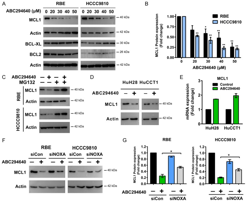
ABC294640 induces degradation of pro-survival protein MCL1. A, B. Western immunoblotting analysis of MCL1, BCL-XL and BCL2 protein levels in RBE and HCCC9810 cells treated with different concentrations of ABC294640 for 24 h. MCL1 protein level following ABC294640 treatment for 24 h was quantified using Image J. Quantitative analysis from three independent experiments (one-way ANOVA with a Turkey post hoc test; data are shown as mean ± SEM; *P < 0.05, **P < 0.01) are shown. C. RBE and HCCC9810 cells treated with no drug control buffer or Proteasome inhibitor MG132 (200 nM) for 2 h, followed by treatment with 50 μM of ABC294640 for additional 12 h. Whole cell lysate was prepared and analyzed for MCL1 expression by Western immunoblotting. Result shown represents 2-3 independent experiments. D. Western immunoblotting analysis of MCL1 protein levels in HuH28 and HuCCT1 cells treated with 50 μM of ABC294640 for 24 h. Result shown represents 3 independent experiments. E. Real-time qPCR analysis of MCL1 mRNA levels in HuH28 and HuCCT1 cells treated with 50 μM of ABC294640 for 24 h. Data are shown as mean ± SEM from 3 independent results. F. Cells were transfected with 50 nM Control siRNA (siCon) or NOXA siRNA (siNOXA) for 48 h and then treated with 50 μM ABC294640 for 24 h. MCL1 protein level was analyzed by Western immunoblotting. Result shown represents 2-3 independent experiments. G. MCL1 protein level of previous experiment was quantified using Image J. Quantitative analysis from 2-3 independent experiments (one-way ANOVA with a Turkey post hoc test; data are shown as mean ± SEM; *P < 0.05) are shown.
