Figure 7.

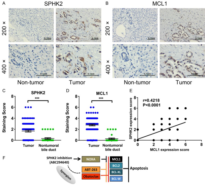
SPHK2 and MCL1 are overexpressed in cholangiocarcinoma and has a positive correlation. A, B. Immunohistochemical staining with anti-SPHK2 and anti-MCL1 antibodies were performed on 89 human cholangiocarcinoma tissues and 31 adjacent non-tumor tissues. Representative images were shown (200× and 400×). C, D. Semi-quantitative analysis of immunohistochemical staining showed that SPHK2 and MCL1 protein expression were upregulated in cholangiocarcinoma in the tissue microarray. Mann-Whitney test were used to compare two groups. ***P < 0.001. E. The expression level of SPHK2 showed a significant positive correlation with MCL1 expression in the tissue microarray. The r and P values were determined by Spearman correlation analysis. F. Schematic representation of the mechanisms of action of ABC294640 in cholangiocarcinoma cells.
