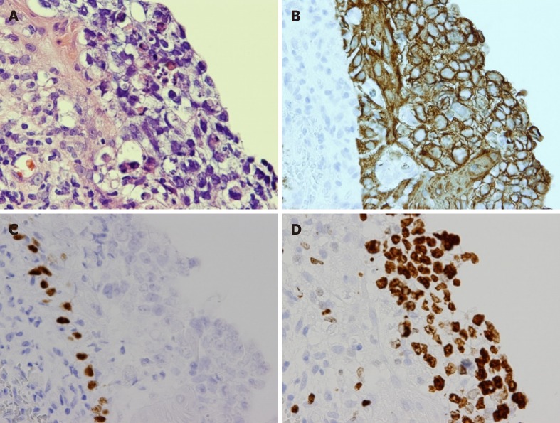
Figure 3.
Pathological findings in case 1. A: Pleomorphic tumor cells with hyperchromatic nuclei, clear cytoplasm and poorly-defined cell borders were seen (hematoxylin and eosin, × 400); B: Immunohistochemically, the tumor cells were positive for AE1/3; C: The tumor cells were negative for p40; D: A high expression of Ki67 was also noted.

