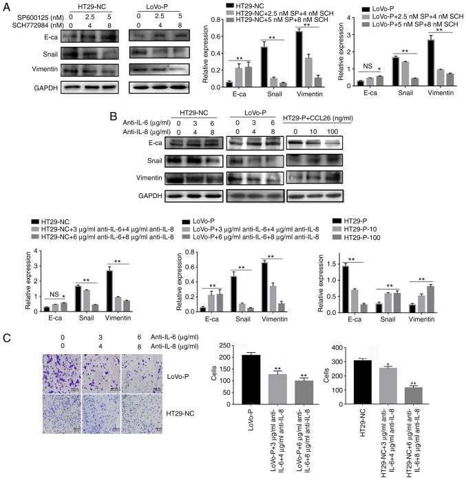
Figure 5.
IL-6 and IL-8 induced epithelial-mesenchymal transition in CRC cells, which was regulated by MAPK and PRL-3. (A) After neutralizing the MAPK pathways in TAMs, the levels of E-cadherin, Snail and Vimentin in colorectal cancer cells were examined using western blot assays. (B) IL-6 (3 and 6 µg/ml) and IL-8 (4 and 8 µg/ml) antibodies, and the addition of CCL26, were used and the levels of E-cadherin, Snail and Vimentin in CRC cells were examined using western blot assays. (C) Invasion was evaluated by Matrigel invasion assays after blocking IL-6 (3 and 6 µg/ml) and IL-8 (4 and 8 µg/ml) *P<0.05, **P<0.01 vs. appropriate controls, NS, not significant. PRL-3, phosphatase of regenerating liver-3; MAPK, mitogen-activated protein kinase; NC, negative control; LoVo-P, LoVo PRL-3 overexpression; HT29-P, HT29 PRL-3 knockdown; E-ca, E-cadherin; SP, SP600125; SCH, SCH772984; IL, interleukin; CCL26, chemokine ligand 26.

