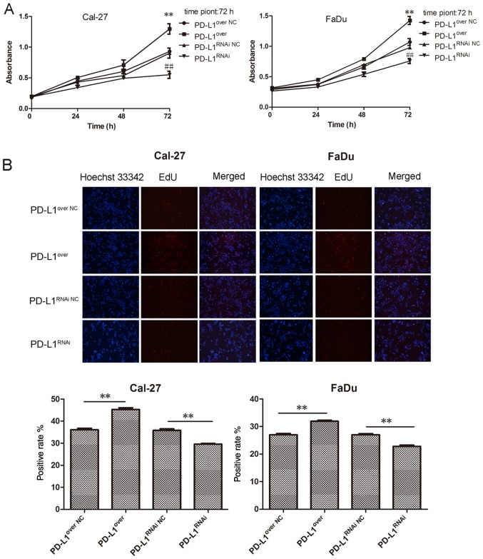
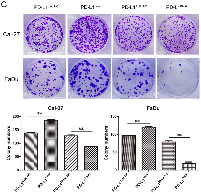
Figure 3.
PD-L1 overexpression promotes the proliferation of HNSCC cell lines. (A) HNSCC cell proliferation was detected with Cell Counting Kit-8 assays, **P<0.01 vs. PD-L1over NC, ##P<0.01 vs. PD-L1RNAi NC. (B) HNSCC cells were labeled with Cell-Light™ EdU (red), and nuclei were stained by Hoechst 33342 (blue). Histograms represent the positive rate of EdU staining. PD-L1 overexpression promotes the proliferation of HNSCC cell lines. (C) Colony formation assay. Cell colony numbers (>50 cells/unit) were counted. Data were expressed as the mean ± standard deviation. **P<0.01. PD-L1, programmed death-ligand 1; HNSCC, head and neck squamous cell carcinoma; PD-L1over, PD-L1-overexpressing; PD-L1RNAi, PD-L1 knockdown; NC, negative control.


