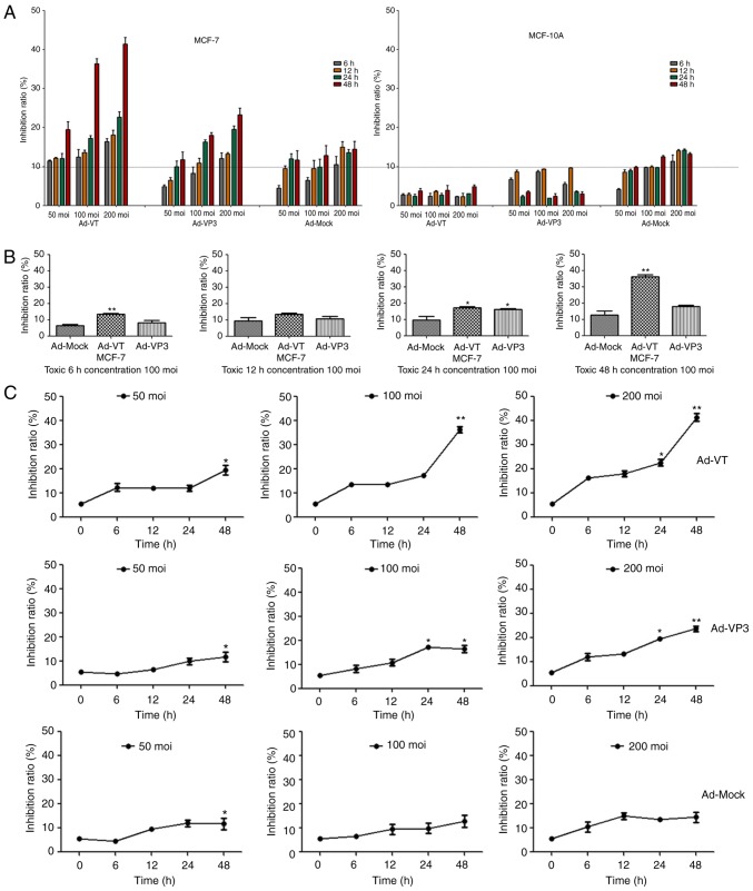
Figure 2.
Recombinant adenoviruses inhibited the proliferation of MCF-7 cells. (A) MCF-10A and MCF-7 cell viability was determined using the WST-1 assay post-infection with various concentrations of Ad-VT, Ad-VP3 and Ad-MOCK (50, 100, and 200 MOI) for 6, 12, 24 and 48 h. (B) MCF-7 cell viability was determined using WST-1 assay after post-infection with Ad-VT, Ad-VP3 and Ad-MOCK (100 MOI) for various durations (6, 12, 24 and 48 h). Data are presented as the means ± standard deviation, representative of three independent experiments (n=3). *P<0.05, **P<0.01, compared with Ad-MOCK. (C) MCF-7 cell viability was determined using the WST-1 assay post-infection with various MOIs of Ad-VT, Ad-VP3 and Ad-MOCK for different durations (6, 12, 24 and 48 h). Data are presented as the means ± standard deviation, representative of three independent experiments (n=3). *P<0.05, **P<0.01, compared with the control (0 h). Ad-VP3, Ad-Apoptin; Ad-VT, Ad-Apoptin-hTERTp-E1a; MOI, multiplicity of infection.

