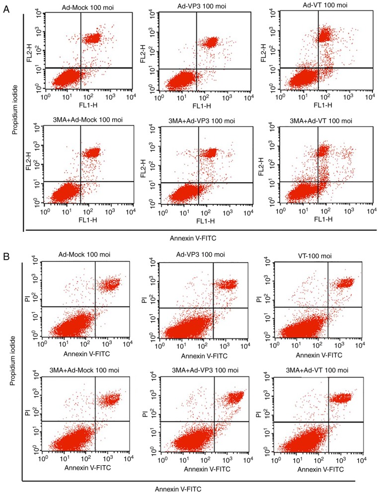
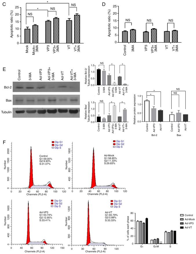
Figure 3.
Analysis of cell death and cell cycle distribution of MCF-7 cells. (A) Scatter plot of Annexin V-FITC/PI staining, which was used to evaluate apoptosis of MCF-7 cells infected with Ad-VT and Ad-VP3 (100 MOI), in the presence or absence 3-MA for 24 h. (B) Scatter plot of Annexin V-FITC/PI staining, which was used to evaluate apoptosis of MCF-10A cells infected with Ad-VT and Ad-VP3 (100 MOI), in the presence or absence of 3-MA for 24 h. Analysis of cell death and cell cycle distribution of MCF-7 cells. (C) Apoptotic ratio, using Annexin V-FITC/PI staining, which was used to evaluate apoptosis of MCF-7 cells infected with Ad-VT and Ad-VP3 (100 MOI), in the presence or absence 3-MA for 24 h. (D) Apoptotic ratio, using Annexin V-FITC/PI staining, to evaluate apoptosis of MCF-10A cells infected with Ad-VT and Ad-VP3 (100 MOI), in the presence or absence of 3-MA for 24 h. (E) Western blotting of MCF-7 cell extracts for Bcl-2 and Bax. MCF-7 cells were infected with Ad-VT and Ad-VP3 (100 MOI) alone, or, after 2 h treatment with 3-MA, Ad-VT and Ad-VP3 was added to the cells for 12 h. (F) Flow cytometric analysis of cell cycle distribution of MCF-7 cells, illustrating the effects of infection with Ad-VT, Ad-VP3 and Ad-MOCK (100 MOI) for 24 h. Data are presented as the means ± standard deviation, representative of three independent experiments (n=3). *P<0.05, **P<0.01, compared with Ad-MOCK. Ad-VP3, Ad-Apoptin; Ad-VT, Ad-Apoptin-hTERTp-E1a; Bax, Bcl-2-associated X protein; Bcl-2, B-cell lymphoma 2; FITC, fluorescein isothiocyanate; MOI, multiplicity of infection; PI, propidium iodide.


