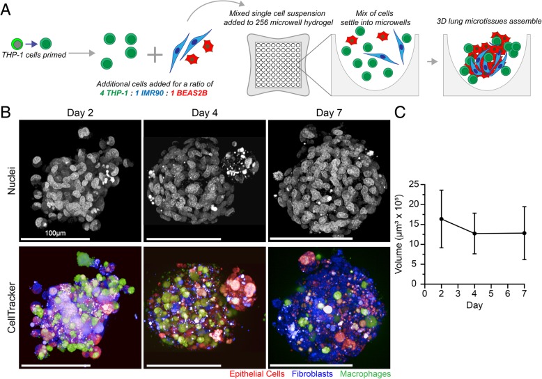
Fig. 1.
Assembly of 3D human lung microtissues. a 3D lung microtissues are plated from a single-cell suspension of three cell lines (BEAS-2B, IMR-90, and THP-1) into a 256-microwell hydrogel. The single cell suspension is seeded into each hydrogel, settles by gravity into the microwells, and cells self-assemble to form microtissues. b Individual cells labeled with fluorescent CellTracker dyes were imaged using confocal microscopy (shown as maximum intensity projections). Scale bar = 100 μm. c Three-dimensional image analysis was used to quantify microtissue volume after 2, 4, and 7 days. Microtissue volume was calculated by averaging the volumes of 142, 177, and 96 microtissues on days 2, 4, 7, respectively. Changes in microtissue volume over time were not statistically significant

