
Fig. 6.
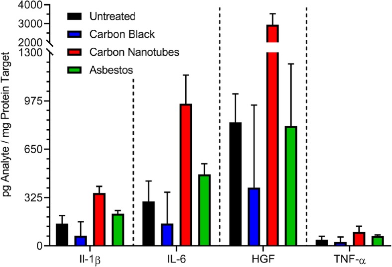
Cytokine protein expression in 3D microtissues exposed to test nanomaterials. Microtissue lysates were analyzed for expression of cytokines (IL-1β, IL-6, TNF-α) or the growth factor, HGF, following exposure to 10 μg/mL of carbon black, carbon nanotubes, or asbestos fibers for 4 days. Mean analyte contents per mg of total protein in the lysate were quantified using Luminex bead assays as described in Materials and Methods. The mean is based on duplicate experiments