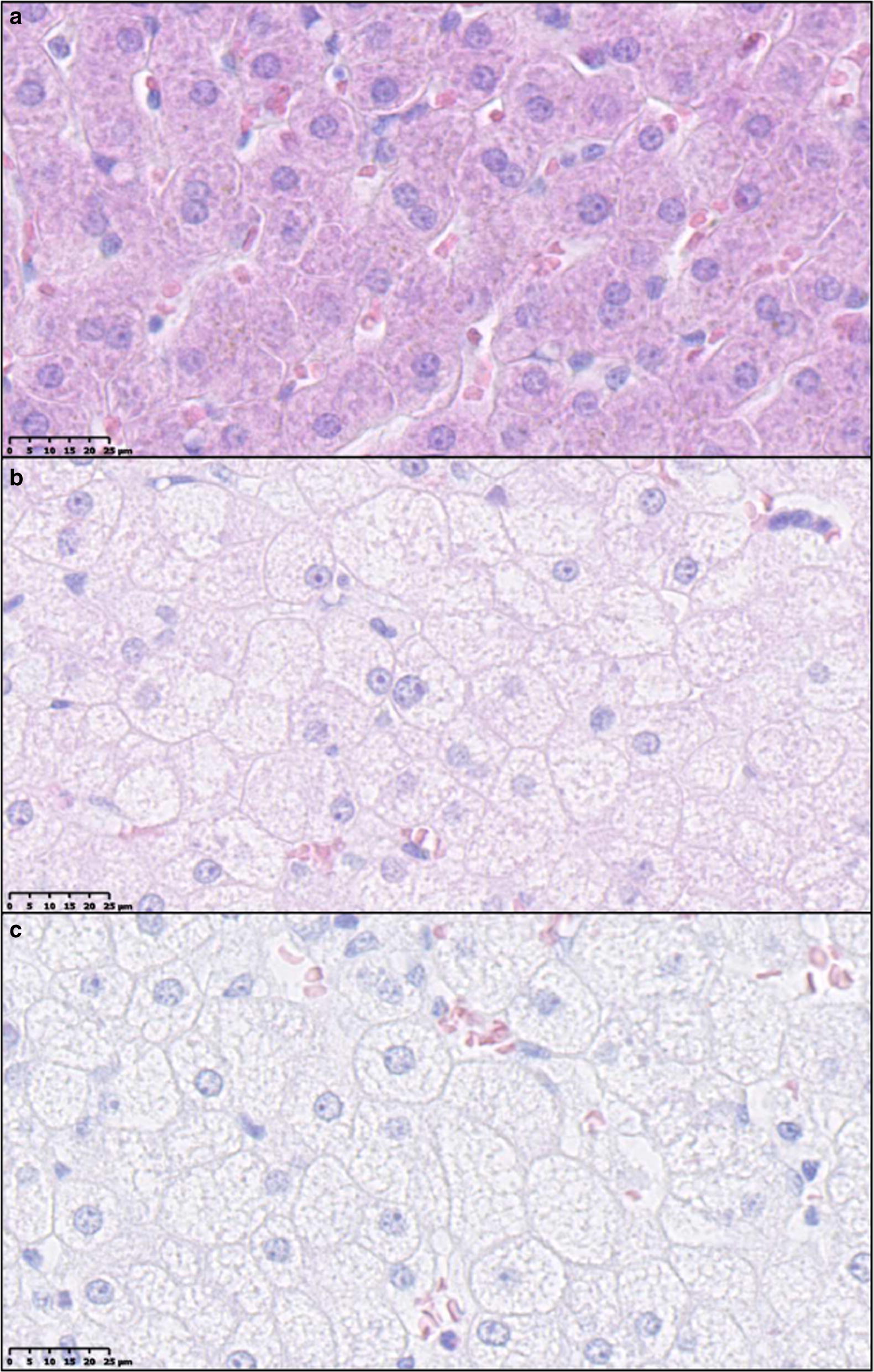
Fig. 5.

Examples of cytoplasmic alterations in hepatocytes characterized by hepatocytes with pale, granular appearance. a Normal hepatocytes from lean control animal (SD). b, c Hepatocytes with cytoplasmic alterations both from animals fed high fat/fructose/cholesterol diet (FFC). Scale bar 25 µm. Hematoxylin and eosin staining
