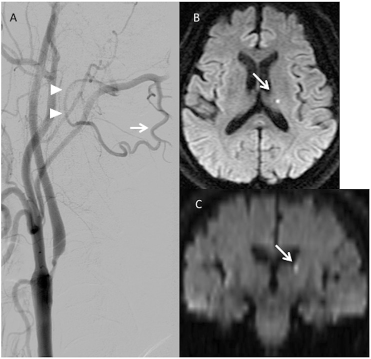
Figure 1.
(a) Patient 1 has a large descending muscular branch (arrow) of the occipital artery, which anastomoses with the vertebral artery (arrowhead) between the atlas and axis. (b, c) Axial and coronal sections of postoperative diffusion-weighted imaging show a small, high-intensity spot in the left lateral ventricle wall above the thalamus (arrow).

