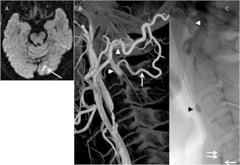
Figure 2.
(a) Preoperative diffusion-weighted imaging shows cerebral infarction (arrow) on the left occipital lobe in patient 6. (b) Three-dimensional rotational angiography shows severe internal carotid artery (ICA) stenosis and a large visible occipital artery (OA)–vertebral artery (VA) anastomosis on the left side. The OA muscular branch (white arrow) anastomoses with the extracranial VA (white arrowheads). (c) A double protection method is applied in the carotid artery stenting procedure. Both 8-Fr (double arrow) and 5-Fr (arrow) guiding catheters are placed into the common carotid artery together. A Percusurge guardwire (black arrowhead) is placed through the 5-Fr catheter into the external carotid artery and Angioguard XP (white arrowhead) is passed through the 8-Fr catheter in the distal ICA.

