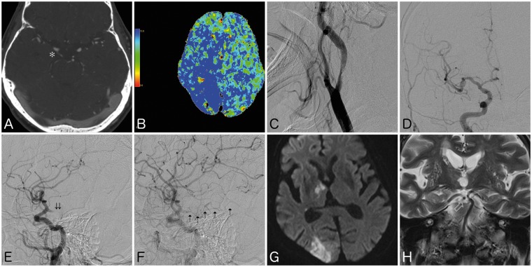
Figure 2.
(a) Computed tomography (CT) angiogram showing intraluminal thrombus located completely within a right fetal posterior cerebral artery (FPCA) (asterisk). (b) CT perfusion scan showing elevated mean transit time involving the posterior right frontal, right temporal, right parietal, and right occipital lobes. (c) Digital subtraction angiogram (DSA) of cervical right internal carotid artery showing severe stenosis with concomitant intraluminal thrombus. (d), (e) DSA in frontal and lateral views showing patency of traditional anterior circulation, with acute occlusion of FPCA (double arrows). (f) Lateral view in late arterial phase showing intralumal thrombus in FPCA with delayed opacification of P3 segments (dashed arrows). (g) Follow-up magnetic resonance image showing moderate infarct of the right thalamus and occipital lobe, with (h) coronal T2 sequence showing patent recanalization of the FPCA. Note the absence of ipsilateral P1 segment.

