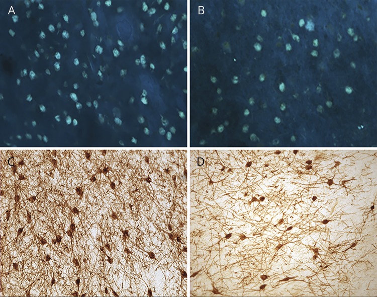
Figure 1. Substantial accumulation of tangles and loss of Ch4 neuronal group of the nucleus basalis of Meynert (nbM-Ch4) neurons in primary progressive aphasia with Alzheimer disease pathology (PPA-AD).
(A) A substantial density of Thioflavin-S-positive tangles was present in the left nbM-Ch4 neurons. (B) A matching section in the right hemisphere shows fewer nbM-Ch4 tangles. (C) Immunostaining for the p75 low affinity neurotrophin receptor (p75LNTR) reveals a dense collection of nbM-Ch4 neurons in control participants, shown here in the intermediate sector of nbM-Ch4 in the left hemisphere. (D) The number of p75LNTR immunoreactive nbM-Ch4 neurons in PPA-AD displays a substantial decrease, shown here also in the intermediate sector of nbM-Ch4 in the left hemisphere. Magnification ×10.

