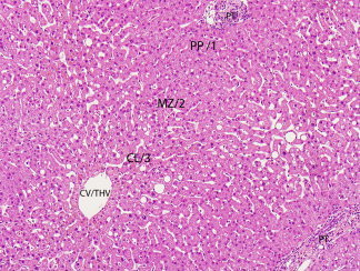
Figure 1.

This photomicrograph illustrates the basic microscopic architecture, structural relationships, and terminology used to describe the microanatomy of the liver. The hepatocellular cords are mostly one or two layers thick and are divided into three zones: 1, 2, and 3 of the acinus, and periportal (PP), mid‐ (MZ), and centrilobular (CL) zones of the lobule. Blood flows from the portal tract (PT) to the central vein, also referred to as terminal hepatic venule (CV/THV) (hematoxylin and eosin stain, magnification ×100).
