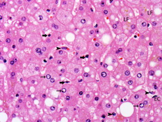
Figure 5.

Sinusoids are lined by endothelial cells (EC) and Kupffer cells (arrowheads). Stellate (Ito) cells (thick arrows) are present in the space of Disse. Clear glycogenated nuclei (thin arrows) and lipofuscin pigment (LF) are also present. Hepatocytes show steatosis, with accumulation of small and large droplets of fat (hematoxylin and eosin stain, magnification ×500).
