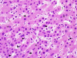
Figure 6.

Area of regenerative change, characterized by mostly smaller hepatocytes (H) that are arranged in cords two or more cells thick, separated by sinusoids (S). The space of Disse is expanded (arrows). A few endothelial cells are also seen (arrowheads). (hematoxylin and eosin stain, magnification ×500).
