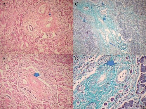
Figure 1.

Histology in noncirrhotic intrahepatic portal hypertension. A combination of photomicrographs depicting the obliterative changes in the portal veins is shown. (A) Thick portal vein with fibrous obliteration of lumen (hematoxylin and eosin stain). (B) Near complete fibrous occlusion of portal venous radicle (hematoxylin and eosin stain). (C) Multiple dilated portal venous radicles in a portal tract (Masson's trichrome stain). (D) Markedly occluded thick walled portal venous radicle (Masson's trichrome stain).
