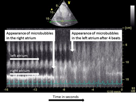
Figure 1.

Contrast enhanced echocardiography in a patient with severe HPS. The figure illustrates the time‐dependent appearence of the contrast agent in the right atrium and four heartbeats thereafter in the left atrium using the M‐mode technique. Abbreviations: M‐mode, motion mode.
