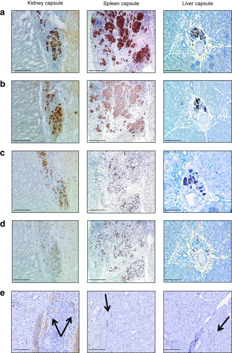
Fig. 2.
Histological analysis of porcine islet transplants. Graft biopsies were sectioned and stained as described in Methods. (a) Porcine islet allografts under the kidney, spleen or liver capsule 120 days after transplantation showed strong staining for chromogranin A. (b–d) Approximately 80% of islet cells showed strong insulin (b), glucagon (c) and somatostatin staining (d). (e) The intensity of CD31 endothelial staining was similar in all tissue types. Scale bars, 400 μm (for figures under the Kidney and Spleen columns) and 200 μm (for figures under the Liver capsule column). Black arrows indicate the site of CD31-positive endothelial staining

