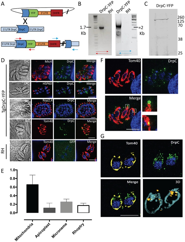
Fig 3. Endogenous tagging of TgDrpC reveals a mitochondria localisation.

(A) Strategy for the endogenous tagging of TgDrpC with YFP in RHΔku80 parasite strain: The ligation-independent cloning vector LIC-YFP was integrated at the 3’ end of TgDrpC, through a single-crossover mechanism. (B) PCR analyses using the primers indicated in (A) confirms the integration of the tagging construct at 5’ and 3’ ends. (C) Western blot analysis using α-GFP antibody on the TgDrpC-YFP clonal line verifies the expected protein size of ≈160 kDa. (D) Immunofluorescence images showing the co-localisation between TgDrpC and the indicated organelles (Mic4, micronemes; HSP60, apicoplast; Rop2-4, rhoptries; Tom40, mitochondrion). (E) Quantification of colocalisation between TgDrpC and the indicated organelles; the Manders’ coefficient (average of n>20 values) is reported in the y axis. (F) TgDrpC puncta (green) have different shapes and sizes, varying from spirals to smaller dots on the membrane; 3-D reconstruction (G) confirms that the vast majority of TgDrpC aggregates are in close proximity with mitochondria. Scale bar: 5 μm.
