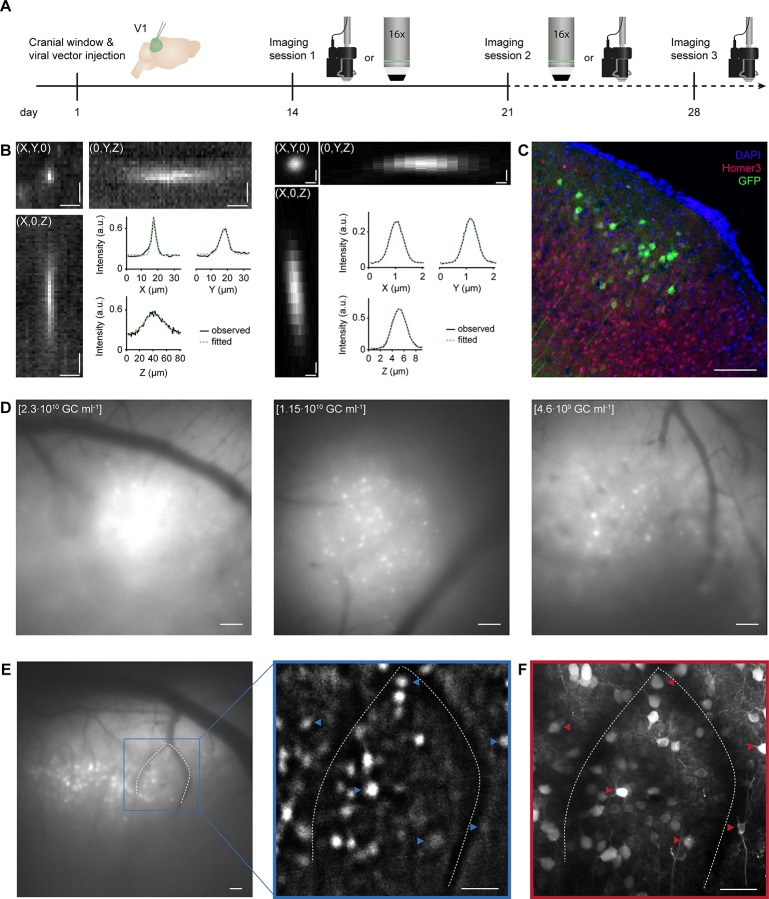
Fig 1. Matching neurons in images acquired with a miniaturized microscope and a two-photon microscope.
A. Experimental timeline in days. Day 1: Cranial window implant and viral vector injection into visual cortex layer 2/3. Days 14 and 21: One miniature and one two-photon microscopy imaging session on either day (order was counterbalanced across animals). Day 28: Optional second miniature microscopy session. Icons at day 14, 21 and 28 illustrate a miniature microscope and a 16x two-photon microscope objective. B. Left: Projections of a three-dimensional stack of observed fluorescence from a sub-resolution fluorescent microbead acquired using a miniaturized microscope. Right: As in Left, but acquired using a two-photon microscope. Insets depict observed (black solid line) and Gaussian-curve fitted (green dashed line) fluorescence intensity along each axis (X, Y and Z) separately. C. Immunohistochemical labeling of GCaMP6s-expressing excitatory layer 2/3 neurons (injection titer of 1.15·1010 GC ml-1). D. Example miniaturized microscopy images of V1 injected with different viral vector titers (left: 2.3·1010 GC ml-1; middle: 1.15·1010 GC ml-1; right: 4.6·109 GC ml-1). E. Left: Miniaturized microscopy image prior to processing. Right: Magnified image after background-subtraction. Blood vessels (dotted lines) assist in matching neurons between microscopes (see panel F; examples of matched neurons are indicated with arrowheads). F. A collapsed volume as imaged with the two-photon microscope (100 planes, 1 μm spacing, projection along the axial axis). Scale bars, 10 μm (B, Left), 0.5 μm (B, Right), 100 μm (C,D), and 50 μm (E,F).

