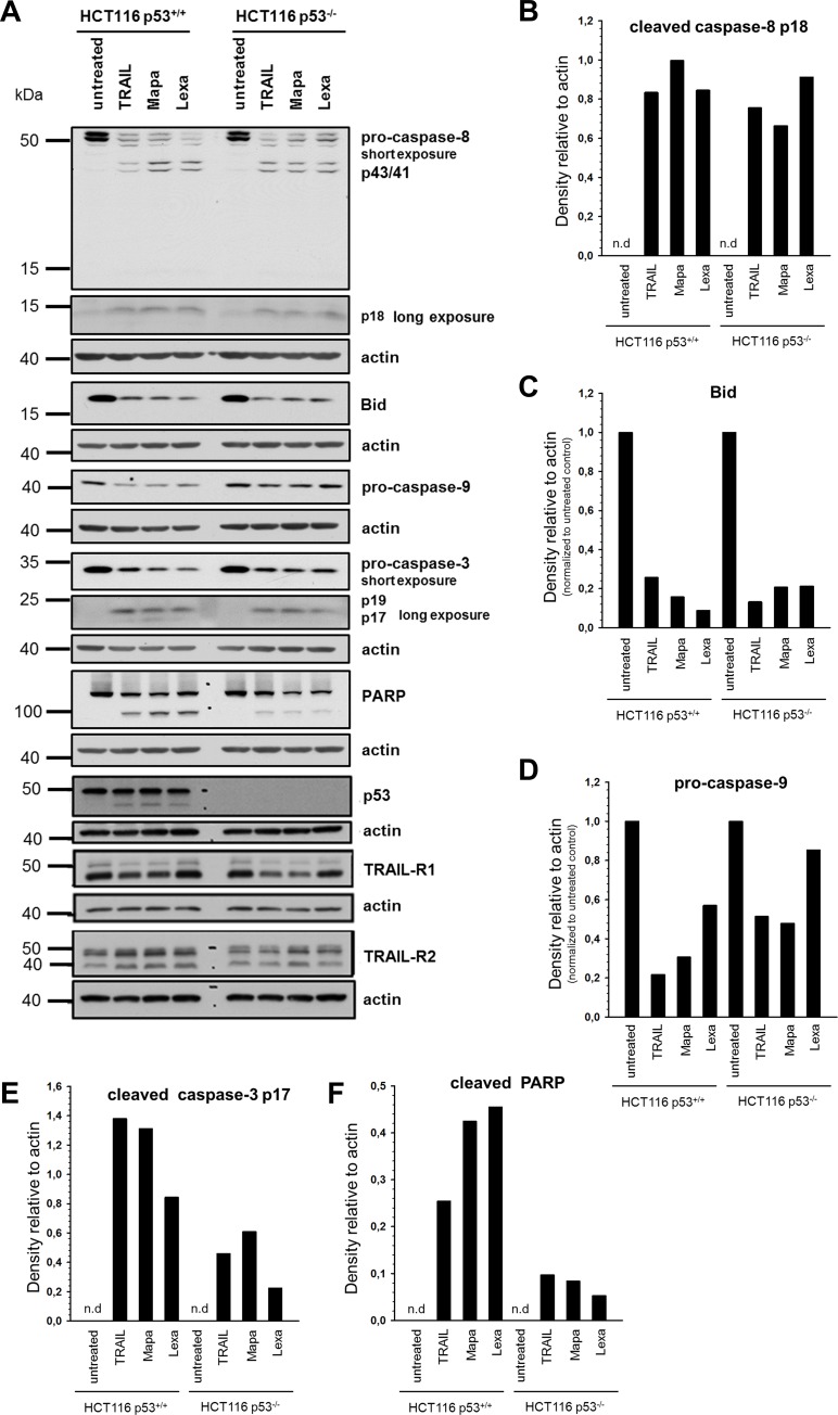
Fig 3. Impact of p53 on TRAIL receptor-induced apoptotic signal transduction pathways in HCT116 cells.
HCT116 p53+/+ and HCT116 p53-/- cells were stimulated with TRAIL (100 ng/ml), Mapatumumab (10 μg/ml) or Lexatumumab (10 μg/ml) for 24 h and the cleavage of pro-caspase-8, Bid, pro-caspase-9, pro-caspase-3 and PARP-1, as markers of different steps of the apoptotic signaling pathway and TRAIL-R1/R2 and p53 were analyzed in whole cell lysates by Western blot (A). Blots are shown for one representative experiment out of three performed. Western blot bands of caspase-8 p18, Bid, pro-caspase-9, caspase-3 p17 and cleaved PARP-1 were analyzed by densitometry (B-F). Intensity of each band was normalized to the respective actin.

