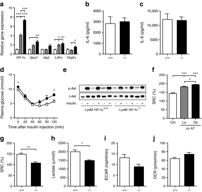
Fig. 6.
Myeloid-specific absence of HIF-1α does not affect adipose tissue inflammation in HFD-fed mice. (a) Fold change expression of Hif-1α and its target genes in BMDMs held in L929-conditioned medium (white bars) or exposed to 100 mg lean adipose tissue explant (grey bars) or obese adipose tissue explant (black bars) for 3 days. Starting quantities were used for normalisation against 36b4. (b, c) IL-6 production by epidydimal adipose tissue isolated from LysM Hif-1α+/+ (+/+) or LysM Hif-1α−/− (−/−) mice, either unstimulated (b) or stimulated with 10 ng/ml LPS for 24 h (c). (d) Glucose measured in plasma of LysM Hif-1α+/+ (white circles) or LysM Hif-1α−/− (black circles) mice upon the injection of insulin at 0 min. (e) Total Akt (t-Akt) and p-Akt protein levels in epidydimal adipose tissue from LysM Hif-1α+/+ or LysM Hif-1α−/− mice unstimulated (−) or stimulated with insulin (+) for 20 min. (f) SRC as percentage increase from basal OCR in BMDMs in 5% (vol./vol.) L929 (Ctrl) or upon 3 days of co-culture (co AT) with 100 mg adipose tissue isolated from lean (Le) or obese (Ob) mice. (g) SRC as percentage increase from basal OCR in BMDMs isolated from LysM Hif-1α+/+ (+/+) or LysM Hif-1α−/− (−/−) mice. (h) Lactate secretion over 24 h by BMDMs isolated from LysM Hif-1α+/+ (+/+) or LysM Hif-1α−/− (−/−) mice. (i, j) Basal ECAR (i) and OCR (j) in BMDMs from LysM Hif-1α+/+ (+/+) or LysM Hif-1α−/− (−/−) mice. Basal ECAR were lower in BMDMs of LysM Hif-1α−/− vs LysM Hif-1α+/+ mice (although the difference did not reach statistical significance, p < 0.051). For the in vivo study, n = 8 animals per genotype/diet were included. n = 3 for all in vitro experiments. Data are presented as means ± SEM. *p < 0.05, **p < 0.01 and ***p < 0.001

