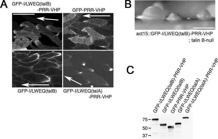
Fig 4.
Sub-cellular localizations of C-terminal actin-binding domains of talin A and talin B. (A) Confocal images of streaming talin B-null cells expressing GFP-I/LWEQ(talB)-PRR-VHP, GFP-I/LWEQ(talB), or GFP-PRR-VHP and talin A-null cells expressing GFP-I/LWEQ(talA)-PRR-VHP. Arrows indicate the direction of migration. GFP-PRR-VHP and GFP-I/LWEQ(talA)-PRR-VHP were also observed in wild-type cells (shown in S2B Fig). (B) Arrested mounds formed by talin B-null cells expressing GFP-I/LWEQ(talB)-PRR-VHP. (C) Immuno-blot analysis using whole cell lysates and an anti-GFP antibody. Marker sizes (kDa) are indicated on the left side of the blot. Scale bars: 10 μm (A) and 200 μm (B).

