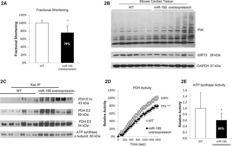
Figure 2. Hyperacetylation and reduced enzymatic activities in miR-195 overexpressing mice.
2A. Echocardiographic analysis showed decreased fractional shortening (p<0.05) in miR-195 transgenic mice compared with WT littermates. Mice were monitored by echocardiography for development of failure at age of 8–10 weeks. 2B. Increased protein acetylation and decreased SIRT3 expression in miR-195 transgenic mice. Whole cell lysates were prepared from heart tissue of WT or miR-195 overexpression mice. Global acetylation level and SIRT3 expression were analyzed by western blotting. GAPDH was used as loading control. 2C. Induced acetylation in PDH complex and ATP synthase α subunit in miR-195 transgenic mice. Whole cell lysates were prepared from heart tissue of WT or miR-195 overexpression mice and subjected to IP assay using anti-Kac. Equivalent amounts of the pellets (IP) were resolved by SDS/PAGE and proteins were detected by immunoblotting. 2D. miR-195 overexpression mice showed a 21% decrease in PDH activity. Errors represent the SD derived from three independent experiments and p<0.01. 2E. miR-195 overexpression mice showed a 40% decrease in ATP synthase activity. Errors represent the SD derived from three independent experiments and p<0.05. * represents p<0.05, ** represents p<0.01.

