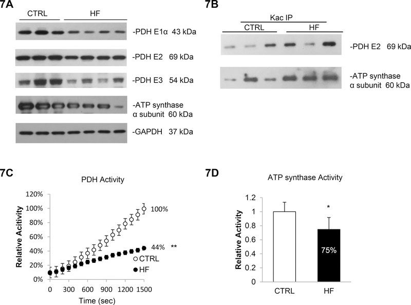
Figure 7. Increased acetylation and impaired metabolism in failing myocardium.
7A. PDH complex subunits and ATP synthase α subunit were less expressed in failing myocardium. Whole cell lysate prepared from normal or HF patients’ heart tissues were analyzed by western blotting. GAPDH was used as loading control. 7B. PDH E2 and ATP synthase α subunit were more acetylation in failing myocardium. Cell lysates prepared from cardiac tissues were subjected to IP assay using anti-Kac. Equivalent amounts of the pellets (IP) were analyzed by western blotting as describe above. 10% of the cell lysate used in the IP reaction was shown as input. 7C. PDH activity was 56% reduced in HF patients. Errors represent the SD derived from three independent experiments and p<0.01. 7D. ATP synthase activity was 25% reduced in HF patients. Errors represent the SD derived from three independent experiments and p<0.05. * represents p<0.05, ** represents p<0.01.

