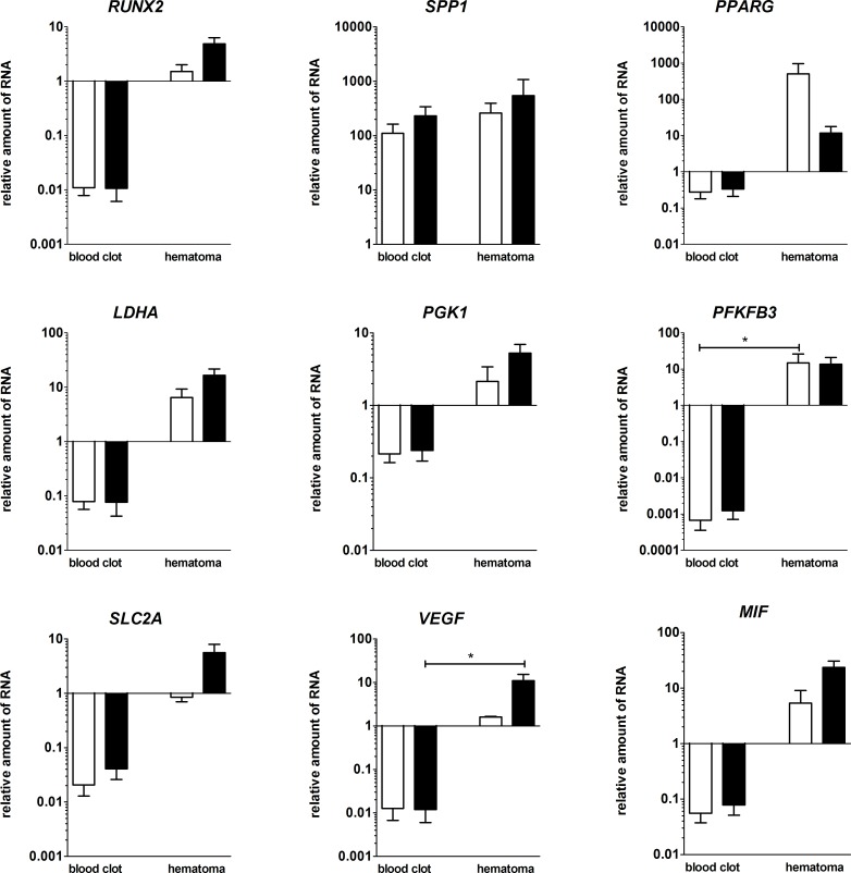
Fig 6. Osteogenic, hypoxia-induced and angiogenic markers were upregulated in the in vitro FH models compared to blood coagulates.
Depicted is the relative RNA-expression of the osteogenic markers RUNX2, SPP1, the adipogenic marker PPARG, the hypoxia induced genes LDHA, PGK1, PFKFB3, SLC2A1 and the angiogenic genes VEGFA and MIF within the FH models and the blood-only coagulates after cultivation in osteogenic differentiation medium for 48 h under either normoxic (white bars) or hypoxic conditions (black bars). All values are normalized to the “housekeeping gene” B2M and to 0 h (mean ± SEM, blood clots: n = 4, FH models: n = 3). Statistical analysis was conducted using Mann-Whitney U-test, *p<0.05.

