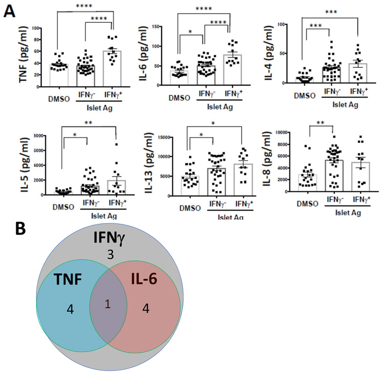
Figure 2. Multiple cytokines are found in the supernatants from library wells from patients with T1D:
(A) Cytokines in the supernatants of IFNγ positive wells (n=12 from 5 subjects with T1D) and IFNγ negative wells (n=32 from 5 subjects with T1D), as well as non-peptide stimulated (DMSO) wells (n=20 from 3 subjects with T1D) were measured with Luminex assay. The levels of IL-2, IL-17, and IL-10 were below the limit of detection. (* p<0.05, ** p<0.01, *** p<0.001, **** p<0.0001, ANOVA with multiple comparisons) The data were combined from 3 independent experiments. (B) Venn diagram is showing the overlap of cytokines from IFNγ positive (n=12 wells from 3 subjects with T1D), TNF positive and IL-6 positive wells. The number of wells are indicated in the graph. The positive was defined by thresholds of mean+3SD of individual cytokines. In addition to IFNγ, which was used to identify the positive wells, other cytokines (TNF, IL-6, IL-4, IL-5, IL-13) were increased in the positive and negative wells but IL-8 was also increased in the IFNγ-cells.

