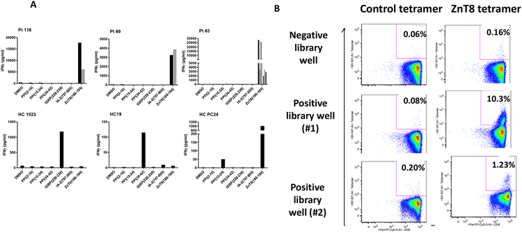
Figure 3. Recognition of ZnT8(186–194) by cells from positive libraries from patients with T1D:
(A) Positive wells from the CD8+ T cell libraries from 4 patients with T1D and 3 HC subjects were further expanded with cytokines and challenged with K562 cells pulsed with each individual peptide used in the original pool. The levels of IFNγ were measured after 6 days. The data are from 11 wells from the 3 patients described in the text (Pt 116, Pt 69, and Pt 63) with T1D and 3 HC subjects (HC1023, 19, PC24). Each graph represents the analysis of positive wells from an individual patient. The bars (black and grey) represent the cytokine responses of different positive wells to the peptides. There were positive responses to ZnT8186–194 in wells from the patients with T1D but responses to IGRP228–236, PPI34–42, PPI15–24, and ZnT8186–194 in the HC subjects. (B) The CD45RO+ cells from one library well without and two library wells with an IFNγ response to the peptide pulsed K562 cells from Pt 63 were expanded in cytokines and stained with tetramers loaded with ZnT8186–194 peptide or control tetramer and analyzed by flow cytometry. The percentages refer to the frequency of tetramer+ cells in the CD8+ gate.

