Figure 6.
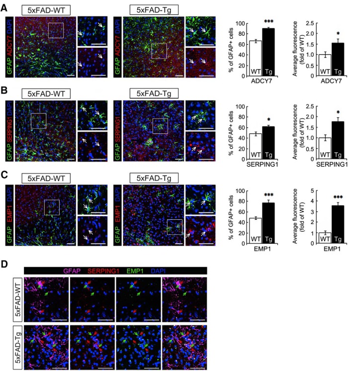
Protein expression of candidate genes in the brain tissues from the 5xFAD Alzheimer’s disease mouse model. Brain tissues collected from 5xFAD mouse models were prepared for immunofluorescence using anti-GFAP (green) and either (A) anti-Adcy7 (B) anti-Serping1, or (C) anti-Emp1. The regions outlined with a square are displayed at higher-magnification on the right, and the arrows point to the same cells for comparison in the fluorescent images. For the astrocytes in the inner layer of cortex (the area near the corpus callosum), the proportion of GFAP+ cells displaying red fluorescence was calculated, and the intensity of red fluorescence overlapping with GFAP signals was measured. Two-tailed unpaired Student’s t tests were used to compare AD samples to the wild-type group. D, Coimmunostaining of cortical brain tissues was performed with anti-GFAP, anti-Serping1, and anti-Emp1. *p < 0.05, **p < 0.01, ***p < 0.001. Scale bars (in A–D), 50 μm. See Extended data Figures 6-1, 6-2, and 6-3.

