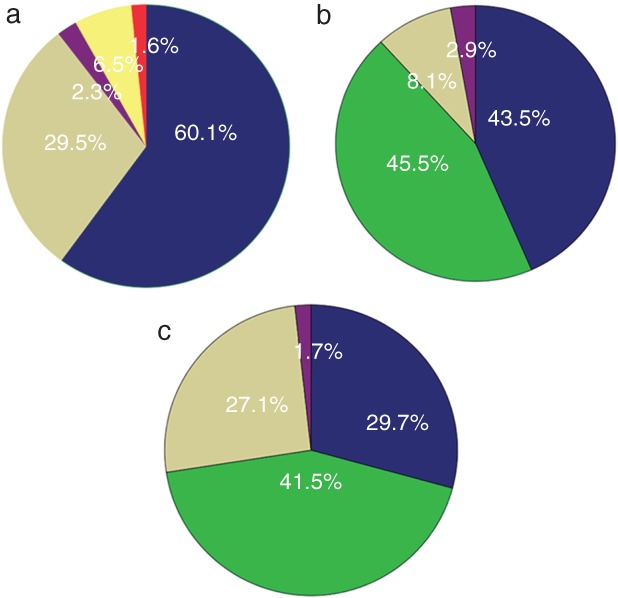
Figure 2.

(a) The proportions of different sampling procedures in patients who underwent re‐biopsy, ( ) CT‐guided PCNB, (
 ) Effusion drainage, (
 ) TBNA/EBUS‐TBNA, (
 ) SLNB and (
 ) other metastasis biopsy. The distribution of results in patients who were tested for EGFR mutation via (b) tissue (c) or plasma. (
 ) Consistent with baseline, (
 ) Mut plus T790M/T790M, (
 ) Wild type, and (
 ) others. “Others” in (b) include three patients with small‐cell lung cancer, two with baseline mutations and c‐MET, one with an exon 20 insert, one with translation from 19del to L858R, one with KRAS and one with ALK. “Others” in (c) include one patient with an exon 20 insert and one with baseline mutations (L858R), S768I, and T790M. CT, computed tomography; EBUS, endobronchial ultrasonography; mut plus T790M, patients harbored a baseline mutation and a T790M mutation when re‐tested; PCNB, percutaneous core needle biopsy; SLNB, superficial lymph node biopsy; TBNA, transbronchial needle aspiration; T790M, patients only harbored a T790M mutation and the baseline sensitive mutation disappeared.
