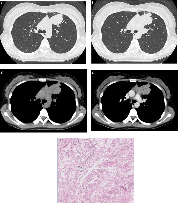
Figure 3.

Computed tomography images of a 30‐year‐old woman with adenocarcinoma with ALK gene rearrangement. (a,b) Lung window images show a solid mass with a spiculated (arrow) and lobulated (arrowhead) margin in the periphery of the left upper lobe. (c,d) Mediastinal window images show the heterogeneous enhancement pattern of the mass. (e) High‐power photomicrograph of the tumor show the acinar predominant subtype (original magnification ×50; hematoxylin and eosin staining).
