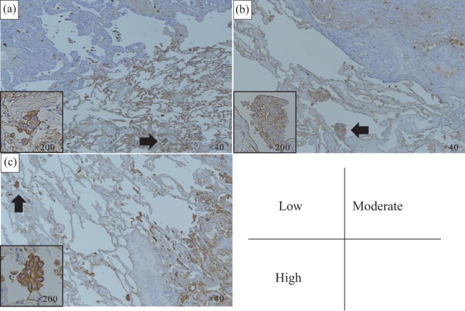
Figure 1.

Immunohistochemical staining of SLX in tumor tissue. The staining was graded as follows: low, one‐third of cells had membranous staining; moderate, one‐third to two‐thirds of cells were stained; and high, ≥ two‐thirds of cells were stained. (a) Low, (b) moderate, and (c) high staining (original magnification ×40). The black arrow indicates tumor spread through alveolar spaces (original magnification ×200).
