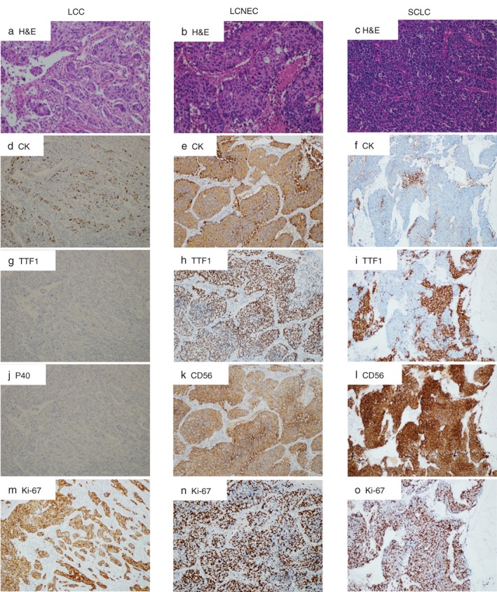
Figure 1.

Morphological and immunohistochemical features of (a) large cell carcinoma (LCC), (b) large cell neuroendocrine carcinoma (LCNEC), and (c) small cell lung cancer (SCLC). Representative examples from each subtype were illustrated. (a) LCC consists of sheets or nests of large polygonal cells. The tumor cell has vesicular nuclei, prominent nucleoli, and moderate amounts of cytoplasm (hematoxylin and eosin [H&E]). Immunohistochemistry (IHC) illustrates dot‐like, cytoplastic staining of (d) CK in tumor cells, (g) negative TTF‐1 and (j) P40, and (m) a high Ki‐67 index. (b) Photomicrograph of LCNEC showing solid nests with multiple rosette‐like structures, with generally large tumor cells with moderate to abundant cytoplasm. Nucleoli are frequent, often prominent (H&E). IHC illustrates (e) a diffuse cytoplasmic pattern of CK expression, (h) positive TTF‐1 with diffuse nuclei staining, (k) positive CD56 in a diffuse membranous staining pattern, and (n) a high Ki‐67 rate. (c) SCLC consists of dense sheets of small cells with scant cytoplasm, finely granular nuclear chromatin, and absent or inconspicuous nucleoli. IHC illustrates (f) a dot‐like, cytoplastic expression pattern of CK, (i) focal TTF‐1 expression, (l) positive CD56 in a membranous staining pattern, and (o) a high Ki‐67 index.
