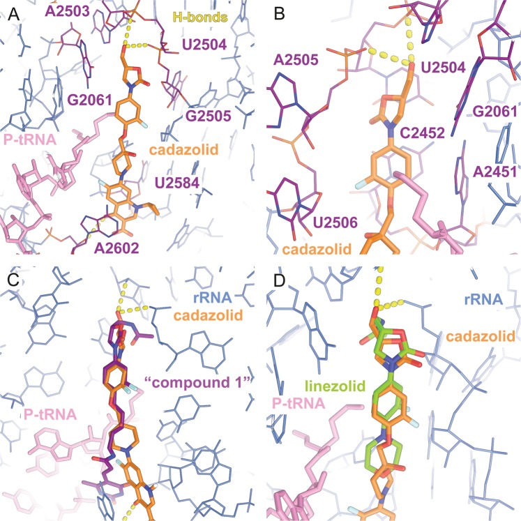
Figure 3.
CDZ bound to the PTC pocket and superposition with structures of two other antibiotics of the oxazolidinone family. (a) Detailed view of CDZ and its interactions with the ribosome. CDZ is shown in orange, the P-site tRNA in pink and the rRNA in blue. The residues forming the pocket where CDZ binds are highlighted in purple. CDZ is within hydrogen-bonding distance of the backbones of A2503 and U2504 (yellow dashes). (b) Close-up view of the A- and B-rings of CDZ. The B-ring is stacking onto residue C2452 while being sandwiched between residues U2506 and A2451. (c) Overlay of CDZ with “compound 1”10,21 or (d) LZD19. The B rings of both oxazolidinones coincide with ring B of CDZ, but the compounds differ in the ring A and C substitutions. “Compound 1”, the tail of which also interacts with A2602, is a hybrid of an oxazolidinone antibiotic and sparsomycin.

